_____________________________________________________________________________________________________________________________________
|
_____________________________________________________________________________________________________________________________________ |
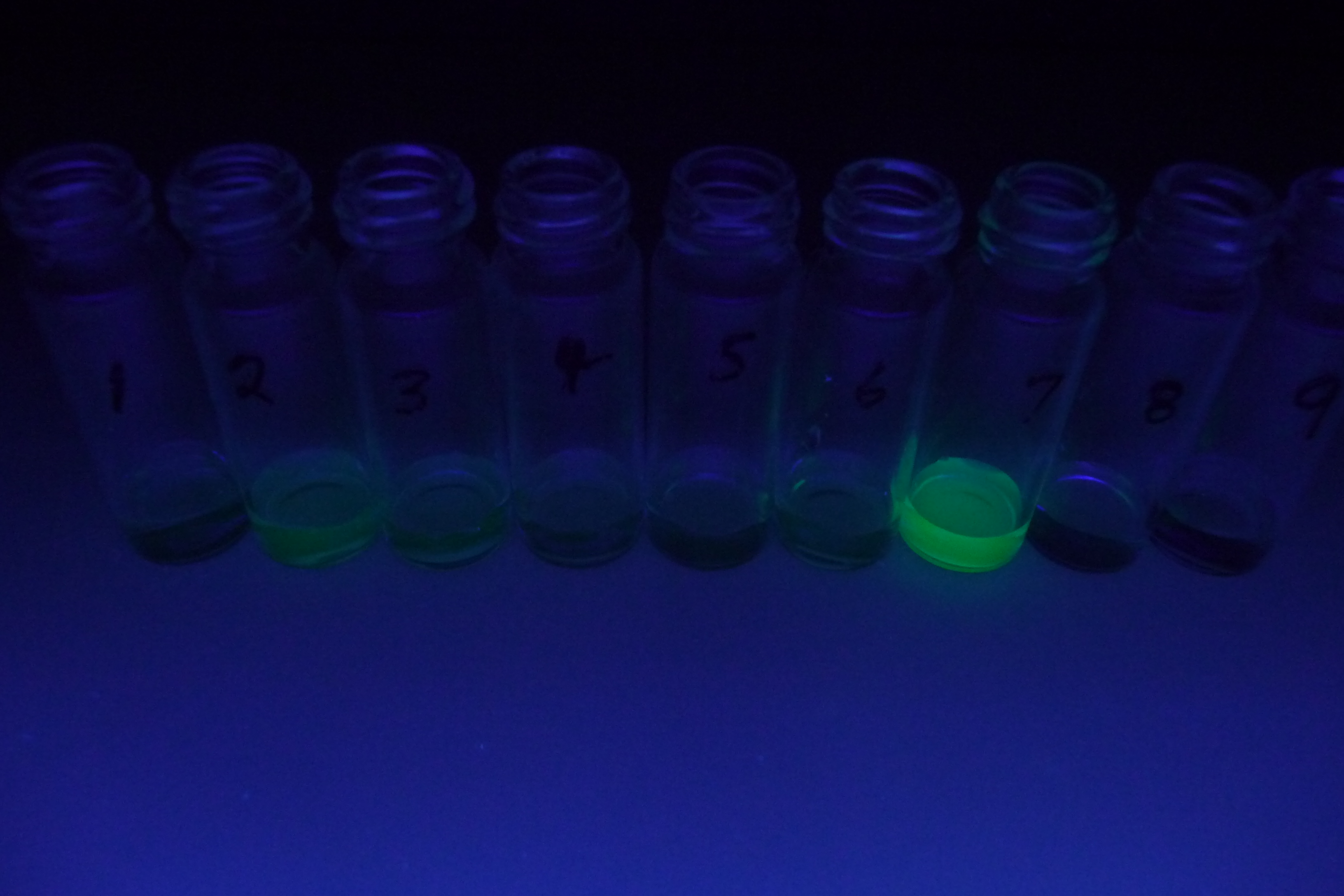
| This image shows a typical analysis of a tagged peptide. Strong green fluorescence can be seen in fraction 7, where the peptide of interest has been separated from impurity peptides, which appear with weaker fluorescence in early fractions. |