Not long after the Green Fluorescent Protein was isolated from jellyfish and characterized, a series of random mutations were performed and found to increase the brightness of GFP fluorescence when the protein is produced at 37 C. The new protein was called cycle 3 GFP. This was a big step forward for making it an important biological tag, since it could be attached to other proteins and grown in animals, and the protein of interest could be followed by visualizing the fluorescence under a microscope. The wild type protein only displays good fluorescence if it is grown at lower temperatures, more similar to the waters of the Pacific Northwest, where the source jellyfish grows.
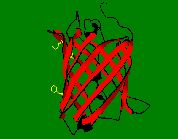
The mutations are F99S, M153T and V163A. This is shorthand for changing phenylalanine at position 99 to serine, methionine at position 153 to threonine, and valine at position 163 to alanine. At first, it was thought the mutations improved the protein's ability to fold at warmer temperatures, but later experiments showed the cycle 3 GFP is much less likely to form non-fluorescent aggregates, most likely due to the removal of hydrophobic residues on its surface. Two of these mutation sites are shown in yellow at right in the wild type protein on the exterior of the molecule. The site of the third mutation (V163T) is also shown in yellow in the interior of the barrel. Because it is in the interior, it most likely does not change aggregation tendency. It is quite clear that amino acid changes that appear to be very minor, which are not close to the chromophore, and which change overall water solubility only slightly, if at all, can have very profound effects on the protein's physical properties.
These experiments are well explained in: Fukuda, H., Arai, M., & Kuwajima, K. (2000). Folding of green fluorescent protein and the cycle3 mutant. Biochemistry, 39(39), 12025-12032.
GFP tryptophan fluorescence
The aromatic amino acids are interesting in part because they display fluorescence. The amino acid with the most intense fluorescence is tryptophan, and it is found the fluorescence is highly dependent on the chemical environment around the side chain indole group. Generally, the fluorescence moves to shorter
wavelengths when the indole is more protected from water solvent, meaning fluorescence provides clues if a particular tryptophan is located on the interior or exterior of a protein.
If the fluorescence emission wavelengths of a molecule (molecule A) overlap with the absorbance wavelengths of a separate molecule (molecule B), and moreover, if molecule B also exhibits fluorescence, then it is possible molecule B fluorescence will be observed when molecule A absorbs light, in a process called Fluorescence Resonance Energy Transfer (FRET). Importantly, the two molecules must be physically close together, at distances from each other which are on the order of a few molecular diameters. FRET efficiency falls of dramatically as the distance increases, with an R^6 dependence, where R is the distance between donor and acceptor.
If one looks at the picture at right, the residue highlighted in blue is GFP's sole tryptophan residue and the one highlighted in green is tryrosine 66. This residue is incorporated into the chromophore which is responsible for the green fluorescence. It turns out tyrosine emission has very good overlap with GFP absorbance, and so when GFP is excited at 280 nm (close to tryptophan absorbance), green emission at 508 nm is observed as if the molecule were being excited at its normal absorbance maximum of 395 nm. Also, tryptophan emission at 320-350 nm is absent. The distance from tryptophan to the chromophore is about 17 Angstrom units.
More below
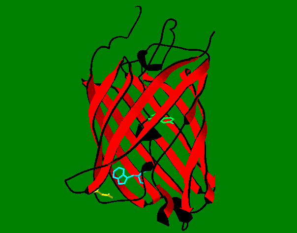
Looking at a bottom view (at right) with the same amino acids highlighted, we see the chromophore as part of the central GFP helix and the tryptophan close to a yellow residue, which is aspartic acid 216.
We have found that the FRET signal observed when GFP is excited at 280 nm can be quenched by certain molecules which gain access to the tryptophan through the bottom of GFP and interact with it such that its energy never makes it to the chromophore, and the green emission is not observed (it is quenched).
Our idea is to change Asp216 to something bigger, such as tyrosine, in hopes of plugging the bottom of GFP and preventing the quenching reaction. This would help prove the quencher is gaining access through the bottom of the molecule, as expected, and not by some other route, such as by changing the hydrogen bonding of the barrel somehow and squeezing through to the tryptophan from the sides. Note that a space filling model of GFP (not shown) makes it clear that many parts of the molecule are completely protected from solvent, such as the chromophore, but that the tryptophan is partially exposed in a fairly deep cleft.
More below
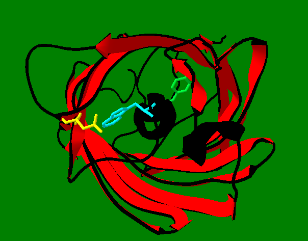
The picture at right shows all 10 GFP tryptophan residues in yellow. They are clearly clustered on one side of the molecule, and in fact it appears close to 1/2 of the protein is tyrosine-free. The phenol side chain of tyrosine has a strong dipole moment, aromatic pi-stacking potential, and a rich free-radical chemistry. Is it possible the unique properties of tyrosine can combine in this cluster to contribute to some of GFP's physical properties? A strong dipole moment for GFP might enhance the polarization of the chromophore and affect its emission wavelength, and this could be influenced by the proper alignment of tyrosine dipole moments.
Two tyrosine residues are found close to the tryptophan. In protein fluorescence, it is often difficult to observe tyrosine fluorescence, and it is often assumed this is due to FRET with tryptophan (if tryptophan is present). In GFP, it is easy to picture tyrosine-to-tryptophan FRET. It is also possible to picture a sort of group effort where any one tyrosine transfers its energy to another tyrosine, where the energy eventually gets to tryptophan (and to the chromophore eventually). This may not be important biologically, since GFP in its natural environment would not experience such short wavelengths of light. But it may help explain why exposing GFP to 280 nm light results in strong emission at 508, and little emission from either tryptophan or tyrosine.
More below
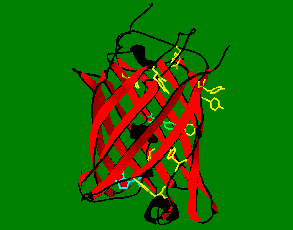
It is fairly easy to change a phenylalanine to tyrosine on strand 11 of GFP. This produces a cluster of 4 tyrosines with their OH groups pointing in close to the same direction and close together in space. Close enough, in fact, it is possible to start thinking about reactions that could link them together. The 4 tyrosines are shown in yellow at right.
There are a number of reasons to consider these reactions:
- If strand 11 is covalently linked to other parts of GFP via a tyrosine, this would enhance the thermal stability of the molecule.
- If the tyrosines are oxidatively linked to make a dityrosine, this would give the molecule new fluorescent properties.
- If the linking chemistry introduces new functionality to the outside of GFP, its chemical properties could be influenced to include enhanced metal binding or hydrophobic character.
More below
One of the observations made during the course of this work is that peptides attached to GFP almost invariably have an effect on the brightness of the fluorescence. That is, each peptide results in a unique phenotype (in the form of brightness) which can be directly linked to the genotype coding for the peptide. This is very puzzling, since the fusion peptide is C-terminal and the GFP molecule should be well on its way to being a fully folded protein by the time the peptide is even expressed. So how can a 20 amino acid peptide tail have such a big effect on brightness? It has been assumed the tail can mimic certain aspects of strand 11 and possibly replace it, which has some sort of an influence on the chromophore, but no evidence to this effect has been obtained.
It turns out that when proteins are synthesized in E. coli, they often need assistance in their folding reactions which is mediated by other proteins called chaperones. An important chaperone is the GroEL/ES system. Since experiments with GFP have shown the protein can be denatured in vitro and then re-natured to its fully fluorescent native state without chaperones, it is assumed GFP folds on its own in vivo, also without the help of chaperones. But its has also been shown that denatured GFP can be captured by GroEL in vitro, where the fluorescence is not restored until the fully functional chaperone is assembled by bringing GroEL in contact with GroES, where the GFP folds properly inside the GroEL/ES cavity. So it is possible GFP folds in E. coli at least in part with the aid of chaperones.
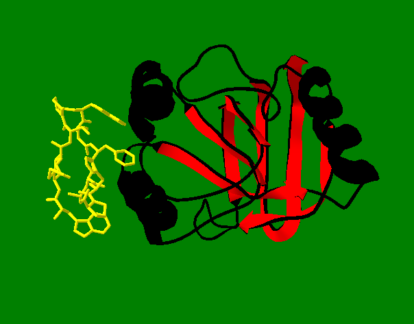
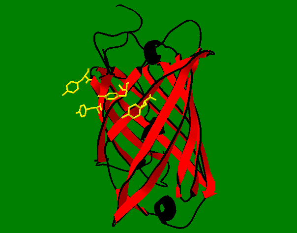
At right is a picture of a peptide (yellow) attached to the binding domain of GroEL. (Fully formed GroEL is much bigger and is worth taking a look at). This is a fairly strong interaction, and in fact helps to map out which parts of GroES bind to GroEL in the process of acting like a chaperone. The yellow peptide is easily attached to GFP using our technology. So, if the GFP-yellow peptide fusion shows a non-fluorescent or dim phenotype, what could this mean? It could mean an unfolded GFP-yellow peptide fusion is sitting in GroEL, where the yellow peptide blocks GroES binding, and the GFP never folds (or folds poorly) and never becomes fluorescent (or becomes weakly fluorescent), because the fully functional GroEL/ES chaperone never forms or forms poorly. We have a number of mutations of the yellow peptide planned that might show this binding can be tuned to be either strong (no fluorescence) or weak (bright fluorescence).